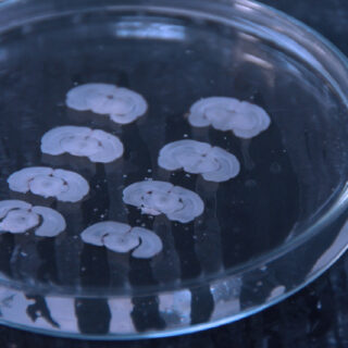
Rodent Brain Slices
Rodent brain slices
We are able to perform electrophysiological recordings on rodent acute brain slices from any brain regions, such as:
- Cortex
- Hippocampus
- Striatum
- Locus Cœruleus
- Thalamus
- Amygdala
- Dorsal Raphe
- Periaqueductal gray Matter
- Cerebellum
- Spinal Cord
- Substancia Nigra
- Dorsal Vagal Nucleus
- Ventral Tegmental Area
- …
PROTOCOLS AVAILABLE
PATCH CLAMP
Passive membrane properties
– Input resistance
– Membrane capacitance
– Access resistance
– Resting Membrane Potential (RMP)
Passive membrane properties
– Input resistance
– Membrane capacitance
– Access resistance
– Resting Membrane Potential (RMP)
PATCH CLAMP
Active membrane properties
– Rheobase
– Spike threshold
– Spike Amplitude
– Firing frequency
– Voltage-gated ion channels
– Ligand gated ion channels
– GPCR modultation
Active membrane properties
– Rheobase
– Spike threshold
– Spike Amplitude
– Firing frequency
– Voltage-gated ion channels
– Ligand gated ion channels
– GPCR modultation
PATCH CLAMP
Synaptic transmission and plasticity
– Evoked responses
– Miniature currents
– Spontaneous currents
Synaptic transmission and plasticity
– Evoked responses
– Miniature currents
– Spontaneous currents
MULTI ELECTRODE ARRAY & HD MEA
– Spontaneous firing activity
– Short term synaptic plasticity
– Long term synaptic plasticity
CALCIUM IMAGING
– Firing activity on small neuronal subpopulation
– Spontaneous firing activity
– Short term synaptic plasticity
– Long term synaptic plasticity
CALCIUM IMAGING
– Firing activity on small neuronal subpopulation
RODENT BRAIN SLICES SAMPLE DATA
MEA – Effect of Carbachol – a cholinergic receptors agonist – on rodent brain slices firing activity.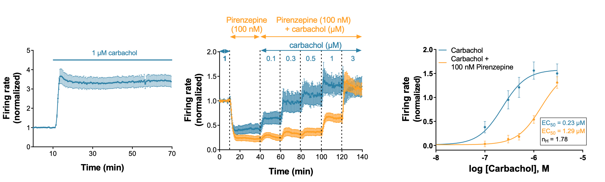
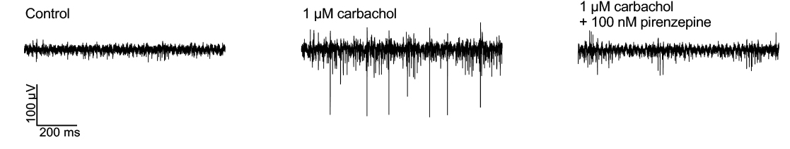
- Carbachol – a cholinergic receptors agonist – strongly increased the firing activity. Carbachol effect stabilized over the 10 first minutes of exposure and then remained steady until the end of the recording session (over 50 minutes).
- Pirenzepine – a selective M1 antagonist – reduced the effect of carbachol (right-shift of the dose-response curve).