Human Brain Slices
Ex vivo human brain slices
Investigating your compound’s activity on human neurons is a key step to help validate early the translation of rodent data to human.
We usually have access to brain tissue coming from two brain regions:
- Neocortex
- Hippocampus
We can perform patch clamp recordings from:
- Pyramidal neurons
- Dentate gyrus granule neurons
- Interneurons
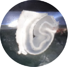
We source our human brain tissue thanks to a partnership with the Bordeaux University Hospital. Human brain tissue is collected on human donors (brain resection from epileptic patients).
PROTOCOLS AVAILABLE
Active membrane properties
– Resting Membrane Potential (RMP)
– Sag
Passive membrane properties
– Rm – Input resistance
– Cm – Membrane capacitance
– Rheobase
– Threshold
– Amplitude
– Halfwidth
– Latency
– fAHP
– Firing rate
– Accommodation
– sAHP
—-
PATCH CLAMP – SYNAPTIC ACTIVITY
Spontaneous EPSC
SAMPLE DATA – Human L2/3 Cortex pyramidal neuron activity
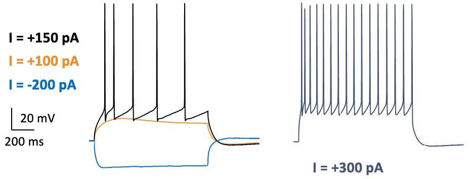
Human L2/3 Cortex pyramidal neuron firing activity
Increasing current intensities are injected into the neuron.
The below graphs shown for several current intensities, the firing activity as a function of time.
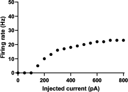
Input / Output curve
Shows the overall firing activity of the L2/3 Cortex pyramidal neuron recorded.
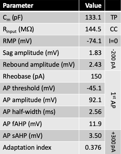
Passive & Active membrane properties of the L2/3 cortex pyramidal neuron
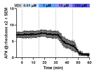
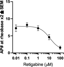
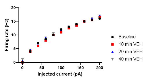
SAMPLE DATA – Kv.7 channel activator Retigabine in cortical pyramidal neurons
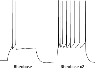
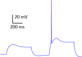
(Left – Vehicule // Right – 100 µM Retigabine)
Dose-inhibiting effect of Retigabine on Pyramidal neuron’s firing at rheobase x2:
