Patch-Clamp
PATCH-CLAMP: SINGLE NEURON ELECTROPHYSIOLOGY
WHAT IS PATCH CLAMP?
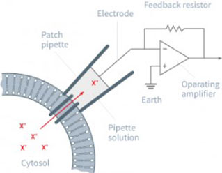
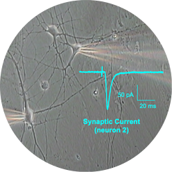
Patch Clamp recordings are performed with borosilicate electrodes of 1-2 μm tip diameter. A pipette containing an electrolyte solution is tightly sealed onto the neuronal membrane and isolates a part of the membrane (patch) electrically.
Currents fluxing through the channels in this patch flow into the pipette and can be recorded by an electrode that is connected to a highly sensitive differential amplifier. The pipette tip makes a giga-ohm seal contact with the neuronal membrane.
Patch Clamp recordings can be performed in different configurations:
- Single channel (as described above)
- Whole-cell (break of the cell membrane and access to the whole membrane conductancies)
WHAT FOR?
This technology provides the highest resolution for electrophysiological recordings from single neuron down to single channel thanks to the different recording configurations (whole-cell, perforated-patch, cell-attached, outside-out).
The Patch Clamp technique is dedicated to the investigation of Mechanism of Action to answer very precise physiological and pharmacological questions.
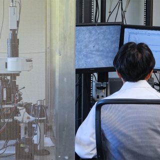
Patch Clamp: a powerful electrophysiology technique
TWO PHYSIOLOGICAL RECORDINGS LEVELS: CELL & SLICE
Patch-clamp recordings is essential to document and understand compounds activity at the single neuron level.
Measurements
- Voltage-gated ion channels
- GPCR modulation of voltage-gated channels
- Ligand-gated ion channels
RECORDING AT THE CELL LEVEL
Rodent Dorsal Root Ganglia neurons
Dorsal Root Ganglion neurons are peripheral sensory neurons essential for Pain preclinical investigations.
We use whole-cell patch clamp recordings on rodent DRG neurons to assess pharmacological agents targeting ion channels.
In addition, we can record DRG neurons from neuropathic rodents to address compound properties specifically developed for neuropathic pains.
This approach provides translational, functional data to assess how molecules act on ion channels and alter their properties.
DRG neurons are a powerful and versatile drug discovery tool for analgesic therapeutics.
Rodent brain neuron cultures
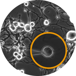
Neuron cultures constitute an easy approach to investigate the effect of compounds on membrane properties (ion channels). This type of assay offers mid-throughput capabilities and small series of compounds profiling.
We can record from cortical neurons and hippocampal neurons.
Cloned channels and receptors
- Stable cell lines
- Transfected cell lines
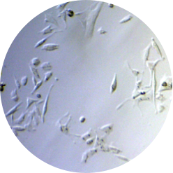
Human iPSC-derived neurons
We are able to physiologically characterise your cells / evaluate your compound’s efficacy on:
- Proprietary iPSCs
- Commercially available iPSCs
Measurements
- Membrane properties
- Voltage-gated ion channels
- Ligand-gated ion channels
- GPCR modulation
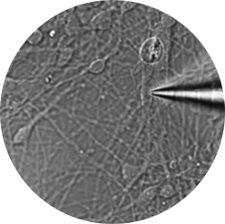
RECORDING AT THE SLICE LEVEL
Rodent brain slice
We have more than 15 years of experience in recording single neuron activity from rodent brain slices. Thanks to state-of-the art slices preparations and protocols designed to fit your needs, we can record from any type of cells to reveal cell-type-specific synaptic and cellular parameters governing neurotransmission.
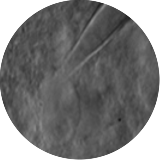
Human brain slice
Thanks to our partnership with neurosurgeons from the CHU of Bordeaux, we have access to human brain tissue resulting from brain resection surgeries.
We are able to perform patch clamp recordings on different cell types:
- Pyramidale neurons
- Dentate gyrus / granule neurons
- Interneurons
We provide different recording types:
- Spontaneous synaptic activity
- Intrinsic properties
- Membrane properties
- Single AP properties
- Trains of APs
