Multi Electrode Array
MULTI-ELECTRODE ARRAY (MEA): NEURONAL NETWORKS ELECTROPHYSIOLOGY
WHAT IS MEA?
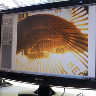
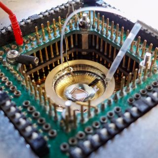
A Multi-Electrode Array is an array made of microscopic metal electrodes (10-30 μm of diameter) distributed on a small surface area (~0.8-6 mm²). They are either regularly distributed or they can be arranged to match closely the spatial organization of the brain region investigated.
These small electrodes (coated with an inert and biocompatible metal) are used for recording electrical signals related to neuronal activities within the slice.
WHAT FOR?
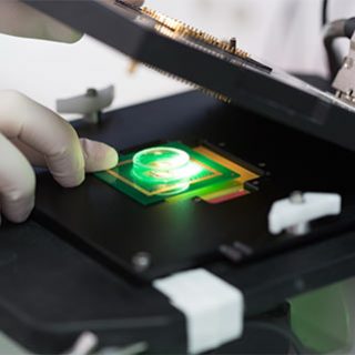
The MEA technology enables parallel and multi-electrode extracellular recordings within a single brain or spinal cord slice. MEA recordings provide an exceptional macroscopic view of neuronal networks.
Thanks to MEA, series of compounds can be evaluated on slices within a couple of days.
WHAT ARE THE ADVANTAGES OF MEA RECORDINGS?
Your compound can be evaluated in vitro under the most physiologically relevant conditions.
Acute brain or spinal cord slices are prepared from rodents (250-500 μm of thickness) and kept alive up to 48 hours.
Neurons and Neuronal Networks display the same physiological properties as in in vivo conditions: all receptors, channels, enzymes, are the native ones with all signalling and regulating pathways being functional.
These slices can be prepared from many different brain regions.
MEA recordings increase the statistical power of the data.
The ability to record multiple evoked-responses in parallel at different electrodes within a single slice and within the same region of interest increases the reliability of the data and their subsequent statistical power.
MEA recordings performed in a single brain or spinal cord slice provide an exceptional macroscopic view of neuronal networks.
Region-specific effects can be readily observed over the MEA electrode surface area. As an example, layer-specific activities can be resolved into stratified structures such as hippocampus or cortex slices.
MEA recordings can facilitate the screening of a series of compounds in parallel, with a remarkably quick turnaround (couple of days).
MEA recordings constitute a well-suited technique for functional series of compound testing as they are much faster than classical glass electrode recordings.